The Center for Diagnostic Imaging at Montefiore Nyack Hospital offers a full range of diagnostic imaging tests, including those explained below.
X-rays use a low-dose beam of radiation to produce an internal image of specific parts of the body. The Center doesn’t require appointments for all X-rays. Exams that do require appointments are:
- Arthrogram
- Barium enema
- Esophogram or barium swallow
- Gallbladder series
- Hystero-salpinogram
- Skeletal survey
- Small bowel study
- Upper GI
- Upper GI with small bowel
- VCUG (adult)
Cardiac computed tomography angiography (CTA) uses special X-rays to focus on the coronary arteries. It allows physicians to see if you have blockages in your heart arteries. To make the vessels in your heart visible on the test, you'll be given an iodine-based contrast material (dye) that will be injected into a vein in your arm. CTA will reveal plaque buildup in your coronary arteries—both calcified “hard” plaque and “soft” plaque, which can be even more dangerous. Having blockage in the coronary artery is one of the main reasons for chest pain and can lead to a heart attack.
CT Scans, sometimes called CAT scans, use advanced X-ray technology to reveal more than what’s possible with a regular X-ray. Our Center has the latest scanner technology available, which produces images of exceptional quality, allowing your doctor to more quickly and accurately provide cardiac imaging, diagnose medical conditions and plan the appropriate treatment. We have the latest generation of CT scanners that produce the lowest radiation dose possible without compromising image quality.
Liver elastography allows doctors to check the liver for fibrosis, a condition that reduces blood flow to and inside the liver. This causes the buildup of scar tissue, which stiffens liver tissue. Left untreated, fibrosis can lead to serious problems, including cirrhosis of the liver, liver cancer and liver failure. But early diagnosis and treatment can reduce or even reverse the effects of fibrosis. There are two types of liver elastography tests—ultrasound elastography and MRE (magnetic resonance elastography—both of which measure the stiffness of liver tissue. Elastography testing may be used in place of a liver biopsy, a more invasive test that involves removing a piece of liver tissue for testing.
Magnetic Resonance Imaging, or MRI, uses magnetism and radio waves to produce clear, internal pictures of the brain, spine, heart or other parts of the body. Sometimes, an intravenous contrast is necessary. Our Center offers the latest MRI technology available, which means faster exam times without compromising image quality.
Cardiac magnetic resonance imaging uses a powerful magnetic field, radio waves and a computer to produce detailed pictures of the structures within and around the heart. Cardiac MRI is used to detect or monitor cardiac disease and to evaluate the heart's anatomy and function.
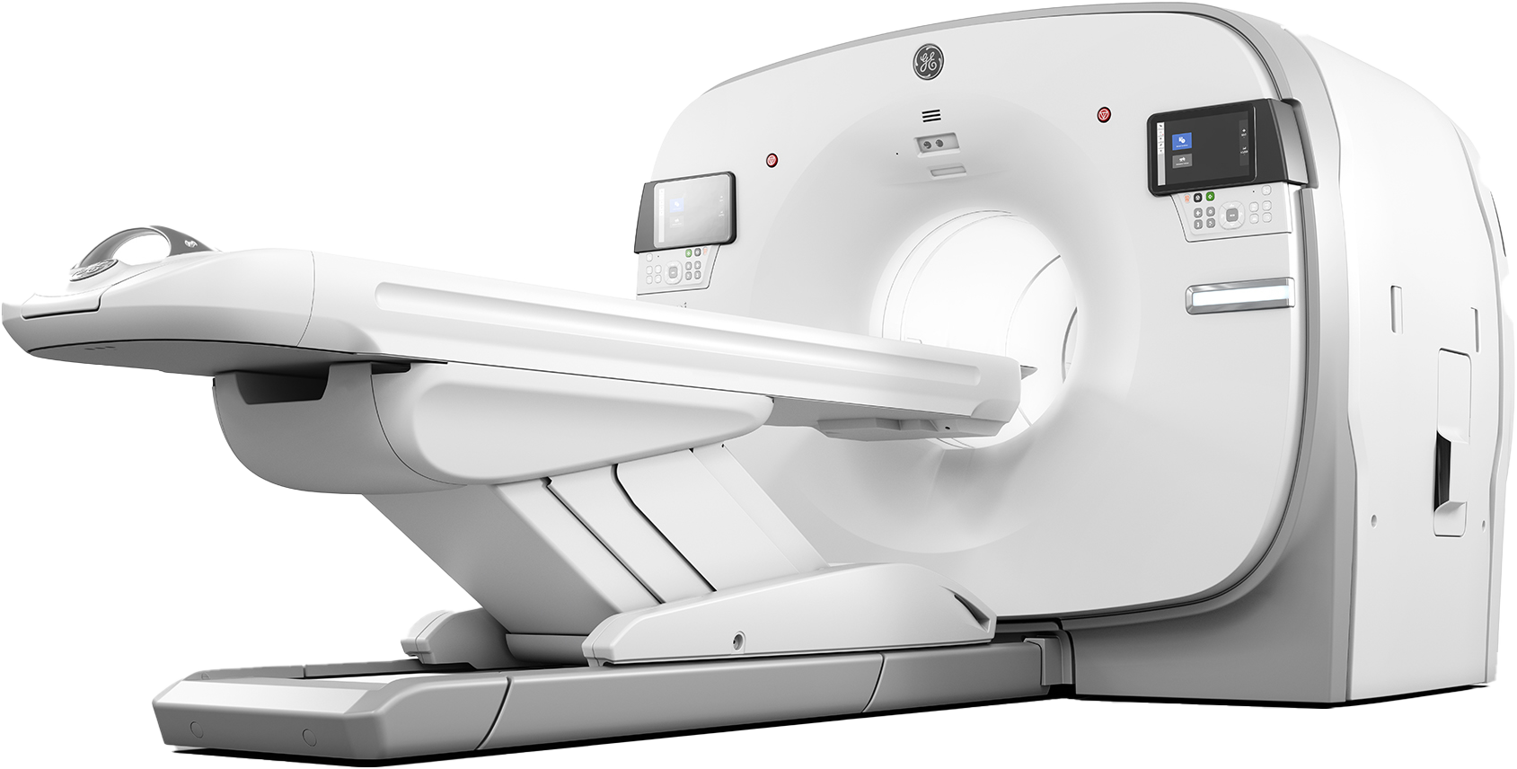
Positron emission tomography (PET) scans look for cancer, check blood flow, or evaluate how an organ is working. They identify the spread or metastases of cancer, which helps determine the most appropriate treatment plan for patients. Radiologists and nuclear medicine specialists analyze and compare the PET scan to CAT scans or Magnetic Resonance imaging results to accurately diagnose patients.
GE Omni Legend PET CT Scanner

Positron emission tomography (PET) scans look for cancer, check blood flow, or evaluate how an organ is working. They identify the spread or metastases of cancer, which helps determine the most appropriate treatment plan for patients. Radiologists and nuclear medicine specialists analyze and compare the PET scan to CAT scans or Magnetic Resonance imaging results to accurately diagnose patients.
- Cardiac, Neurologic, Oncologic and Urologic Studies
- Scan time and injectable isotope reduced up to 30%
Nuclear Medicine exams use small doses of radioactive material to produce internal images of the body. Nuclear medicine differs from other imaging exams in that it reveals how an organ is working, not just what it looks like. Nuclear medicine is also used to detect bone and joint abnormalities, including trauma, fractures, arthritis and tumors. Some common nuclear medicine tests include:
- Bone scan
- Hepatobiliary scan
- Hyperthyroid treatment
- MUGA study
- Nuclear stress test
- Prostascint scan
- Thyroid cancer ablation
- Thyroid scan
Ultrasound uses high-frequency sound waves to capture images. There's no contrast injection or radiation exposure associated with ultrasound, making it an ideal tool to monitor fetal development. Our Center offers ultrasound exams on a variety of areas, including the abdomen, pelvis, thyroid, renal glands and scrotum. Ultrasound can also capture blood flow in the vessels and document moving images of the heart.


 Upcoming Events